Back in May 2017, I saw a postdoc job advert which stated:
“The project aims to develop and build a novel dual-channel circularly-polarised luminescence (CPL) microscope and evaluate its performance throughout live-cell studies of parallel synthesized CPL active responsive Lanthanide(III) complexes.”
I am very glad that I got job, and joined Durham Chemistry as part of a great team in the group of Dr Robert Pal.
First thing for me to do was to develop a spectrometer for CPL light. That work took a bit longer than originally expected, but was published in Nature Communications in 2020. Then, I got waylaid by a combination of a work with collaborators, a successful BBSRC Discovery fellowship application, and the small matter of a global pandemic. We were actually starting to put together the back-end of this CPL-measuring confocal microscope (based upon CPL the rapid spectrometer) when the COVID pandemic closed down laboratory access.
Bizarrely being forced out the lab meant I was able to focus my attention on writing a review on CPL-active security inks for Nature Reviews Chemistry; the rapid CPL spectroscopy technology we had previously developed finally making verifying CPL-active security inks a practical proposition.
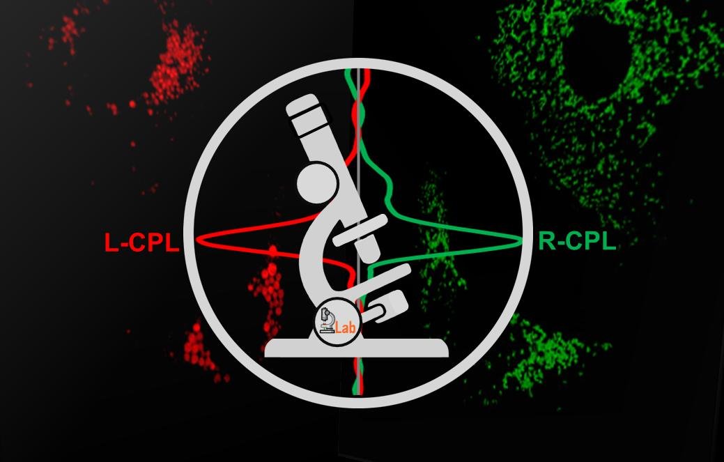
By the time labs had re-opened I was a BBSRC Discovery Fellow, working on upconversion nanosensors. Fortunately we had recruited a brilliant postdoctoral researcher, Dr Patrycja Stachelek to take over from me. Cue some long days in the microscopy lab getting everything carefully aligned, with lots of swearing and beeping of equipment. Patrycja and Rob did a great job developing test targets, carrying out multiphoton CPL measurements, and finally, live-cell imaging of enantiopure samples of luminescent CPL-emitting complexes. Resulting, finally, in January 2022: the world’s first CPL confocal microscope published in Nature Communications.
This microscope is uniquely capable of tracking the left and right handed luminescent molecules as they journey into cells. Our demonstration utilized a complex developed by Parker and Pal groups at Durham, showing that one enantiomer localised to the lysosome and the other enantiomers localised to the mitochondria.
I really look forward to seeing how the technology is exploited going forward. New sub-cellular probes revealing chiral mechanisms behind molecular biology? Micro-patterned CPL-active ultra-secure security inks? Time will tell….
It was a pleasure to be part of this work and this team.
I’m also pleased to see that the work has been featured on several news websites, including:
